 Asbestos Flooring Test Lab Procedures & Photos
Identify Floor Tiles or other Products That Contain Asbestos
- POST a QUESTION or COMMENT On Asbestos Testing & Asbestos Test Labs
Asbestos test lab procedures, photographs, sample preparation, microscopy methods & example lab report: this article provides photos and procedural suggestions used in the forensic laboratory to identify asbestos materials (or probable-asbestos) in flooring and floor tiles.
InspectAPedia tolerates no conflicts of interest. We have no relationship with advertisers, products, or services discussed at this website.
- Daniel Friedman, Publisher/Editor/Author - See WHO ARE WE?
Asbestos Test Procedures: Microscopy to Identify Asphalt-Asbestos or Vinyl-Asbestos Floor Tiles in the Laboratory
 In this article series we provide photographs and descriptive text of asbestos insulation and other asbestos-containing products
to permit identification of definite, probable, or possible asbestos materials in buildings.
[Click to enlarge any image]
Asbestos is safe and legal to remain in homes or public buildings as long as the asbestos materials are in good condition and the asbestos can not be released into the air.
While an expert lab test using polarized light microscopy is necessary to reliably identify the presence and specific type of asbestos fiber, or to identify the presence of asbestos in air or dust samples, many asbestos-containing building products not only are obvious and easy to recognize, but since there were not other look-alike products that were not asbestos, a visual identification of this material can be virtually a certainty in many cases.
On occasion, the original flooring packaging or installation literature may be available for a given home: often an extra box of floor tiles was kept for future repairs.
The vinyl-asbestos floor tile package label information, combined with a simple comparison of tiles in the package with tiles installed in the building may be sound confirmation of asbestos-containing materials.
See Vinyl Asbestos Floor Tile Packaging. Historical information about the dates of flooring installation may also be sufficient to rule in or out the possibility that flooring in a building contains asbestos.
Walter McCrone developed and amply documented the forensic microscopy procedures used to identify asbestos in products, air or dust samples.
Certainly asbestos-certified labs who process large volumes of asbestos samples have developed efficient, high-speed procedures to keep the sample analysis costs down, and surely some of those experts have other tips and ideas for effective processing of floor tile samples besides what we will document here.
However on occasion we need to work with less sample material, or very small asbestos floor tile sample fragments in our laboratory. Here we document and illustrate some suggestions for working with small fragments of vinyl-asbestos floor tiles to obtain material for microscopic examination in the laboratory.
Preparing a Small Floor Tile Fragment for Microscopic Examination for Asbestos
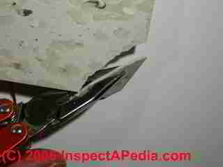
In the lab, following Walter McCrone's procedure for teasing out asbestos particles from solid materials such as this floor tile, we broke a small corner off for further examination by microscope.
Tiles are broken, not cut, in order to expose asbestos fibers for removal, slide preparation, and microscopic examination using transmitted, reflected, and primarily polarized-light central stop diffusion microscopy.
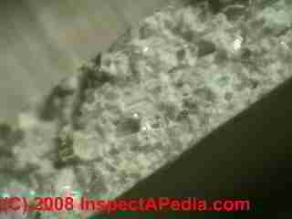
This stereo-microscopic view of the edge of this asbestos-floor tile shows the combination of binder and other silicate materials.
Details about Montgomery Ward vinyl asbestos tile flooring are
at MONTGOMERY WARD ASBESTOS FLOOR TILE IDENTIFICATION.
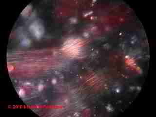
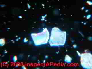
Below (left) is a microphotograph of materials (probably limestone filler) scraped from the broken edge of the Wards vinyl asbestos floor tile shown above. And below (right) is a 1200x magnification photo taken in our laboratory, showing asbestos fibers teased out of the broken edge of a separate sample of floor tile tentatively identified as 2mm x 9"x9" Armstrong Excelon vinyl asbestos flooring ca 1954-1980).
Because many fibers such as fiberglass and asbestos can be almost impossible to detect microscopically, especially in small fragments, unless they are mounted in a proper medium, Cargille certified refractive index liquids (e.g. n=1.550 or n=1.680) are used to mount asbestos-suspect fibers for microscopic examination.
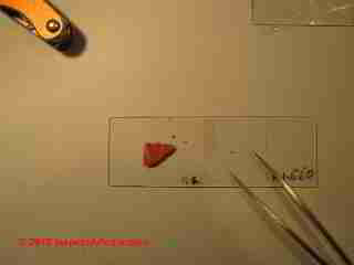
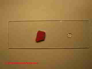 Our photo (left) shows a small fragment of red floor tile (tentatively identified as Armstrong Apache Red) prepared for initial handling.
Although McCrone instructs the technician to "tease out" fibers from the edge of a broken floor tile fragment, the "teasing out process" can be tricky if like the author (DF) you have stubby fingers and the sample is about 1cm square.
We use a very small quantity of fast-setting glue to bond our floor tile fragment to a clean microscope slide.
Watch out: don't use too much glue. Especially "super glue" or "Krazy glue", convenient to work with, may dissolve into the sample fragment, or wet its important broken sample edge if you use too big a glue droplet.
To the right of the floor tile fragment you'll see a drop of clean mounting fluid. We're using Cargille™ N=1.680 in this example. We'll use the droplet to secure fibres we remove from the sample.
Watch out: don't make the droplet too big or you'll waste time looking for your sample fibers under the microscope; don't place the droplet too close to the glued sample or you may overload it with granular debris during fiber removal; don't place the droplet too far away from the sample or you may lose a fiber during transfer from the sample to the drop.
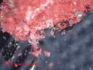

At above left we show our probe under the stereo microscope, as we gently pick away material from the exposed edge of the glued floor tile sample fragment in order to expose a cluster of fibers for further examination.
Our photo at above right shows that we check our mounting fluid droplet for obvious extraneous debris before using it. If the droplet becomes debris-loaded, it's easy to clean the slide and start with a new drop since we've glued down our sample.
Above a fiber cluster has been removed from the sample and carried to the nearby droplet of mounting fluid. We did not try to remove all of the debris from our fiber cluster as we wanted to keep the fiber bundle intact.
Below, we still haven't lost the sample as we further prepare the slide with a cover slip. If we are using the same slide and glued-sample to prepare a sequence of fiber trials at different Cargille liquid values, we note the refractive index on the slide so as to keep our lab data accurate.
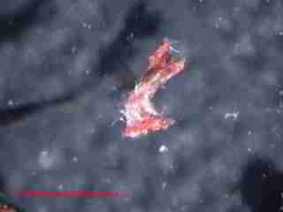
The following six asbestos microscope photos, below show the asbestos fiber cluster from our vinyl asbestos floor tile under magnification and at different lighting conditions.
...
...
Our last photo (below) show mineral fragments from the sample, possibly limestone.
Watch out: very find powdered asbestos was used as a non-fibrous filler in many products.
...
Continue reading at ASBESTOS FLOORING HAZARD REDUCTION or select a topic from the closely-related articles below, or see the complete ARTICLE INDEX.
Or see these
Recommended Articles
- ASBESTOS TEST LABS
- ASBESTOS TESTING SAMPLE COLLECTION
- FLOOR TILE HISTORY & INGREDIENTS for a discussion of the ingredients and production of asbestos-containing flooring.
- ASBESTOS FLOORING IDENTIFICATION for advice on visual identification of vinyl-asbestos floor tiles or flooring products that probably do or don't contain asbestos.
- DUST SAMPLING PROCEDURE
Suggested citation for this web page
ASBESTOS FLOOR TILE LAB PROCEDURES at InspectApedia.com - online encyclopedia of building & environmental inspection, testing, diagnosis, repair, & problem prevention advice.
Or see this
INDEX to RELATED ARTICLES: ARTICLE INDEX to ASBESTOS HAZARDS
Or use the SEARCH BOX found below to Ask a Question or Search InspectApedia
Ask a Question or Search InspectApedia
Try the search box just below, or if you prefer, post a question or comment in the Comments box below and we will respond promptly.
Search the InspectApedia website
Note: appearance of your Comment below may be delayed: if your comment contains an image, photograph, web link, or text that looks to the software as if it might be a web link, your posting will appear after it has been approved by a moderator. Apologies for the delay.
Only one image can be added per comment but you can post as many comments, and therefore images, as you like.
You will not receive a notification when a response to your question has been posted.
Please bookmark this page to make it easy for you to check back for our response.
IF above you see "Comment Form is loading comments..." then COMMENT BOX - countable.ca / bawkbox.com IS NOT WORKING.
In any case you are welcome to send an email directly to us at InspectApedia.com at editor@inspectApedia.com
We'll reply to you directly. Please help us help you by noting, in your email, the URL of the InspectApedia page where you wanted to comment.
Citations & References
In addition to any citations in the article above, a full list is available on request.
- "Asbestos in your home or at work," Forsyth County Environmental Affairs Department, Winston-Salem NC 12/08
- [1] "Asbestos in the Home," U.S. EPA, Exposure Evaluation Division, Office of Toxic Substances, Office of Pesticides and Toxic Substances, U.S. Environmental Protection Agency, Washington,D.C. 20460also see EPA’s epa.gov/asbestos/pubs/ashome.html
- New York State Department of Health, Wadsworth Center, "ELAP Labs Certified for Asbestos", web search 04/16/2011, original source: http://www.wadsworth.org/labcert/elap/asbestos.html. Additional contact information for the Wadsworth Center: David Axelrod Institute Wadsworth Center NYS Department of Health P.O. Box 22002 Albany, New York 12201-2002
- New York State Department of Health Corning Tower Empire State Plaza, Albany, NY 12237 - Public Health Duty Officer Helpline 1-866-881-2809
- Thomas Hauswirth, Managing Member of Beacon Fine Home Inspections, LLC and (in 2007) Vice President, Connecticut Association of Home Inspectors Ph. 860-526-3355 Fax 860-526-2942 beaconinspections@sbcglobal.net 06/07: thanks for photographs of transite asbestos heating ducts
- Gary Randolph, Ounce of Prevention Home Inspection, LLC Buffalo, NY, for attentive reading and editing suggestions. Mr. Randolph can be reached in Buffalo, NY, at (716) 636-3865 or email: gary@ouncehome.com 3/07
- Asbestos Identification, Walter C.McCrone, McCrone Research Institute, Chicago, IL.1987 ISBN 0-904962-11-3. Dr. McCrone literally "wrote the book" on asbestos identification procedures which formed the basis for current work by asbestos identification laboratories.
- Stanton, .F., et al., National Bureau of Standards Special Publication 506: 143-151
- Pott, F., Staub-Reinhalf Luft 38, 486-490 (1978) cited by McCrone
- In addition to citations & references found in this article, see the research citations given at the end of the related articles found at our suggested
CONTINUE READING or RECOMMENDED ARTICLES.
- Carson, Dunlop & Associates Ltd., 120 Carlton Street Suite 407, Toronto ON M5A 4K2. Tel: (416) 964-9415 1-800-268-7070 Email: info@carsondunlop.com. Alan Carson is a past president of ASHI, the American Society of Home Inspectors.
Thanks to Alan Carson and Bob Dunlop, for permission for InspectAPedia to use text excerpts from The HOME REFERENCE BOOK - the Encyclopedia of Homes and to use illustrations from The ILLUSTRATED HOME .
Carson Dunlop Associates provides extensive home inspection education and report writing material. In gratitude we provide links to tsome Carson Dunlop Associates products and services.


