
InspectAPedia® FREE Encyclopedia of Building & Environmental Construction, Diagnosis, Maintenance & Repair |
Question? Just ask us! InspectAPedia
|
 Mold spores under the Microscope
Photo ID Library of common molds found in buildings

- POST a QUESTION or COMMENT about what building mold looks at under the light microscope at magnifications from 10x to 1200x.
What does mold look like under the microscope?
Organized alphabetically by mold name (mold genera/species), these mold spores and their photographs (both on site and under the microscope) have been collected in the U.S., Spain, Mexico, France, as well as in other countries.
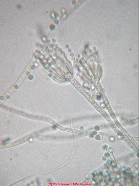
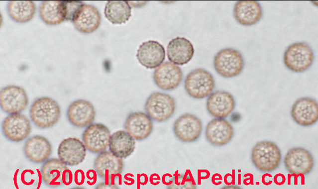
These are aerobiology laboratory photos of mold under the microscope. taken by the author unless otherwise stated. The photo at page top shows Aspergillus sp., fungal spores and condiophores under the microscope at 1200x.
InspectAPedia tolerates no conflicts of interest. We have no relationship with advertisers, products, or services discussed at this website.
- Daniel Friedman, Publisher/Editor/Author - See WHO ARE WE?
MOLD BY MICROSCOPE - What mold looks like under the light microscope

In this article we provide lab photographs of mold under the microscope.
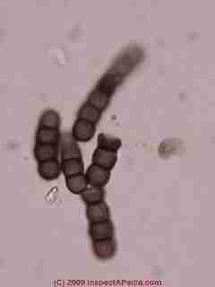
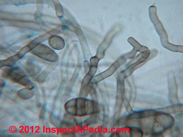
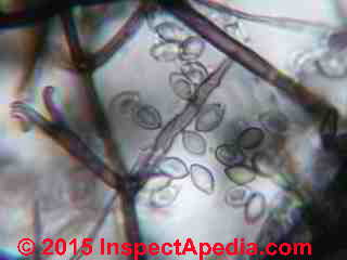
Just above our photo shows chains of of Aureobasidium pullulans.
Other photographs and articles at this website will help you find and recognize problematic toxic or allergenic mold in buildings.
Nearly all of our mold spore photographs shown at this mold spore identification assistance page are from field samples collected in buildings.
What makes these photographs helpful is that they are "real world" examples of mold spore occurrence, including the surrounding debris and sometimes rough growth patterns of mold spores that occur in situ in buildings.
Mold grown in the laboratory or on cultures is often very crisp, beautiful, and perhaps more easy to identify. But the actual physical structures of mold growth for a given genera and species may vary significantly depending on the material on which the mold is growing - its food.
Photographs of mold spores under the microscope shown here are in that sense, more "natural" than those obtained from culture.
See MICROSCOPE DIGITAL PHOTOGRAPHY for advice on how to obtain and organize good photographs through the microscope.
Photographs of Mold Under the Microscope
Mold spore photographs are arranged alphabetically here. For example, if you are looking for what Stachybotrys chartarum spores and growth structures or conidiophores look like under the microscope, just scroll down to the "S" section of our identification photographs of mold under the microscope.
Index to Building Mold Genera/Species in This Document
A - B - C - D - E - F - G - H - I - J - K - L - M - N - O - P - Q - R - S - T - U - V - W - X - Y - Z
A
Acremonium-like Mold Spore Photographs

Here is a laboratory microscopic photograph of Acremonium like mold growth found on wet "fuzzy" basement vinyl flooring and at a water heater leak in Missouri.

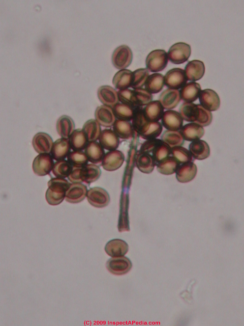
Agrocybe sp. mold spore photographs
Photograph of an Agrocybe spore (see Basidiomycetes). Individual Agrocybe spores are common in outdoor air samples collected with spore traps.
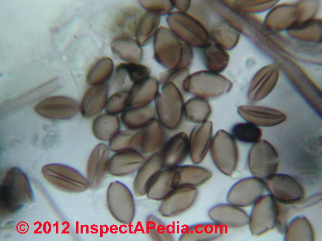
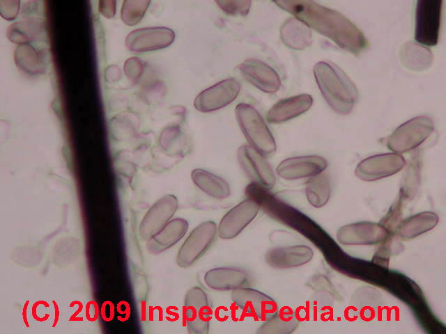
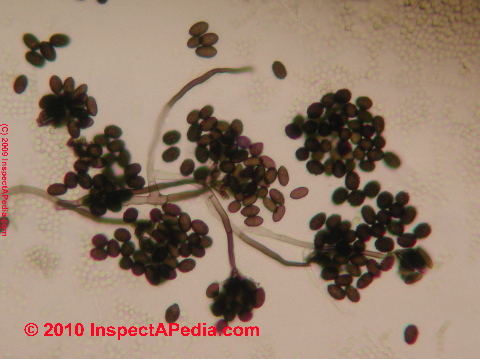
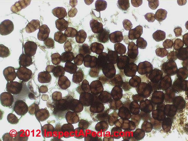
Alternaria sp. mold spore photographs
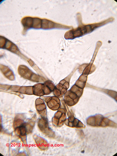
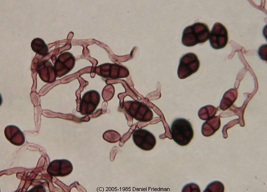
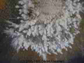
The photograph of Alternaria spores in a cluster (above shows these spores with their proper coloration. The Alternaria sp. photograph below demonstrates the confusion in spore coloration and thus in spore identification that can occur when a novice microscopist relies too heavily on fuchsin stains for spore detection.

Alternaria mold spores are very common in outdoor air and are likely to be found in outdoor air samples and are often found in indoor air samples as well. Growing on a building surface (or in culture) Alternaria sp. will also appear in spore chains (photo at left) and attached to fungal hyphae.
Our Alternaria sp. mold spore chain photo just above also includes skin flakes and at bottom center an Ascomycete.
Comparing the Alternaria spores to the human skin cells and to the smaller Ascomycete you can see that Alternaria fungal spores are quite large among members of the Fifth Kingdom.
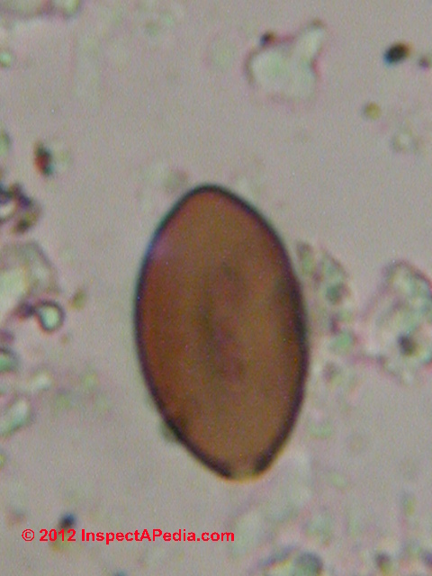
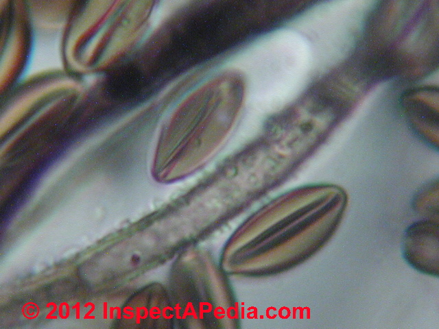
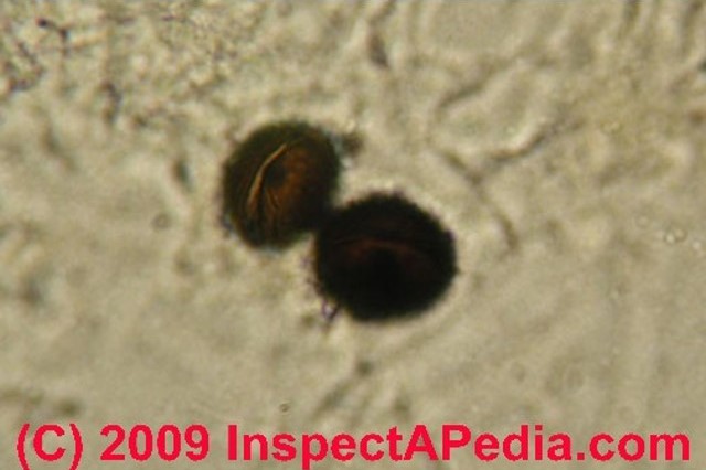
Arthrinium sp. mold spore photographs
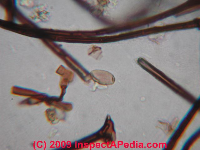
Arthrinium fungal spores (in close-up at 1200x) form group of at least twenty species, some of which are ovate or lemon-shaped. Possible examples of A. phaeospermum are shown below. This fungus is often confused with the ubiquitous Chaetomium sp. fungal spore when the latter is not fully hydrated
. Look for not a fold in the spore (dessicated) but a hyaline band at the junction of the two sides, and look for the birth scar (bottom of the spore at below right) - that's an Arthrinium sp. spore not Chaetomium sp. [Thanks our instructors, mycologists Dr. Harriet Burge and Dr. John Haines.]
Chaetomium sp. is an Ascomycete, born in groups of 8, and without a birth scar as it emerges from a perethicium not by growing on a fungal hypha. And the center fold on Arthrinium will extend pretty much to the ends of the spore.
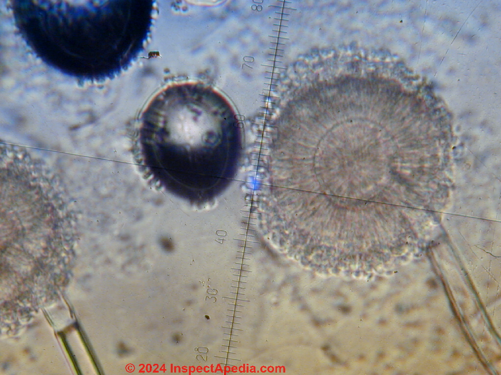
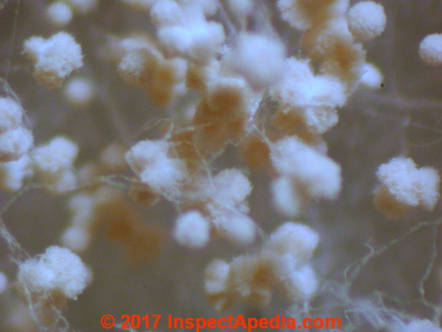
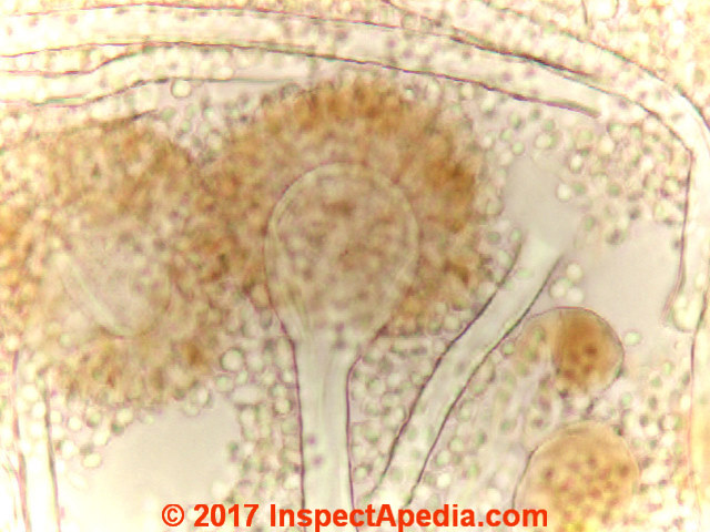
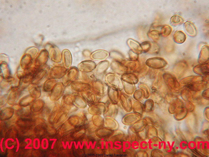
Aspergillus sp. mold spore photographs
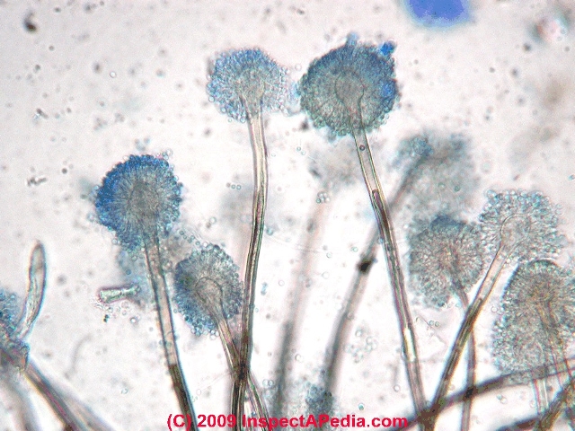
Photographs of Aspergillus sp. mold spores under the microscope Aspergillus niger culture, Penicillium culture, Penicillium spores - Aspergillus and Penicillium spores are difficult to differentiate when they are found in air that you may see them reported in test results as "Pen/Asp".
Watch out: Most Pen/Asp spores are round, hyaline (colorless) and small and lack surface features to aid in their precise identification by microscope when the spores are found alone, or in air samples (and if not in spore chains).
In that case the spores may not even be identified as (potentially harmful) molds and may just be called "amerospores" in the lab report.
But when these spores appear in chains (as that's how they are born) they should not be labeled as "amerospores", and at least some of these airborne spores in the Aspergillus/Penicillium group can be identified from the spore alone, however, such as Aspergillus niger
Also see below our lab photograph of mold spores in this dense surface growth of Aspergillus niger and Aspergillus fumigatus side by side, in the same surface sample, but not quite intermixed.
Below Aspergillus versicolor-like mold growing in culture and under the microscope; note that usually A. versicolor produces very long mold spore chains atop the conidiophore, not seen in these images.
See MOLD APPEARANCE - WHAT MOLD LOOKS LIKE for images of what mold looks like when growing on building surfaces or materials.
Aspergillus penicilloides is not common in the indoor envrionment but may be detected. [Lab photo needed, use the page top or bottom CONTACT link] See this research citing Aspergillus penicilloides in the indoor environment (and the U.S. Space Station):
- Gock, Melissa A., Ailsa D. Hocking, John I. Pitt, and Peter G. Poulos. "Influence of temperature, water activity and pH on growth of some xerophilic fungi." International Journal of Food Microbiology 81, no. 1 (2003): 11-19.
- Meklin, Teija, Tiina Reponen, Craig McKinstry, Seung-Hyun Cho, Sergey A. Grinshpun, Aino Nevalainen, Asko Vepsäläinen, Richard A. Haugland, Grace LeMasters, and Stephen J. Vesper. "Comparison of mold concentrations quantified by MSQPCR in indoor and outdoor air sampled simultaneously." Science of the Total Environment 382, no. 1 (2007): 130-134.
- Twarużek, Magdalena, Ewelina Soszczyńska, Piotr Winiarski, Aleksander Zwierz, and Jan Grajewski. "The occurrence of molds in patients with chronic sinusitis." European archives of oto-rhino-laryngology 271, no. 5 (2014): 1143-1148.
- Vesper, Stephen J., Wing Wong, C. Mike Kuo, and Duane L. Pierson. "Mold species in dust from the International Space Station identified and quantified by mold-specific quantitative PCR." Research in microbiology 159, no. 6 (2008): 432-435.
- Vesper, Stephen, Craig McKinstry, Richard Haugland, Lucas Neas, Edward Hudgens, Brooke Heidenfelder, and Jane Gallagher. "Higher environmental relative moldiness index (ERMI sm) values measured in Detroit homes of severely asthmatic children." Science of the total environment 394, no. 1 (2008): 192-196.
Abstract:
Sieved vacuum bag dust from the homes of 143 children in Detroit was analyzed by mold specific quantitative PCR (MSQPCR) and the Environmental Relative Moldiness Index (ERMIsm) was calculated for each home.
Children living in these homes were grouped as non-asthmatic (n = 83), moderately asthmatic (n = 28) and severely asthmatic (n = 32) based on prescription medication usage for their asthma management (none, occasional and daily, respectively). The mean ERMI for each group of homes was 6.2 for non-asthmatic, 6.3 for moderately asthmatic and 8.2 for severely asthmatic children.
The ERMI values in the homes of severely asthmatic children were significantly greater compared to the non-asthmatics (p = 0.04 in Wilcoxon Rank-sum test). Aspergillus niger and Aspergillus unguis were the primary mold species that distinguished severely asthmatic children's homes and non-asthmatic children's homes (p < 0.05; Wilcoxon Rank-sum test).
The determination of the home's ERMI values may aid in prioritizing home remediation efforts, particularly in those children who are at increased risk for asthma exacerbation. - Wady, Loay, Annicka Bunte, Christina Pehrson, and Lennart Larsson. "Use of gas chromatography-mass spectrometry/solid phase microextraction for the identification of MVOCs from moldy building materials." Journal of microbiological methods 52, no. 3 (2003): 325-332.
Harvard
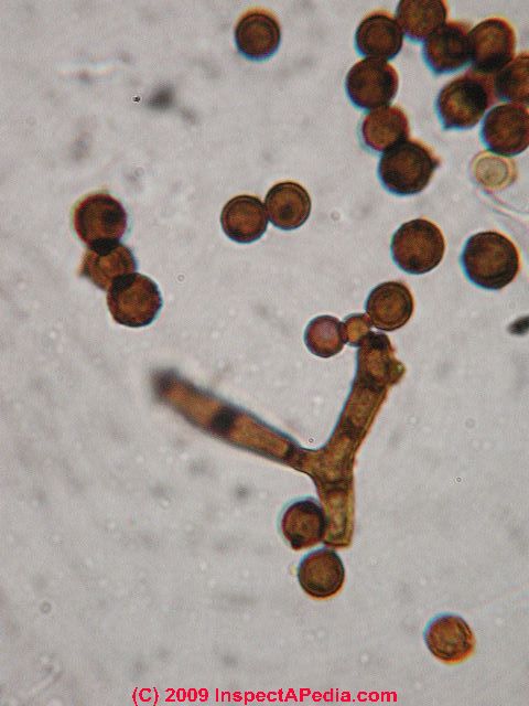
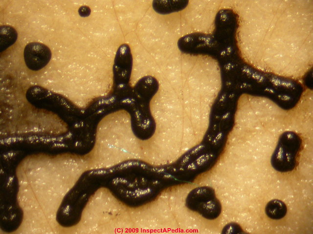
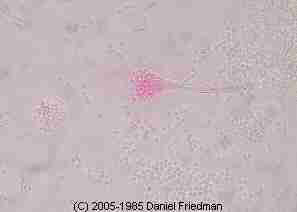
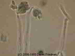
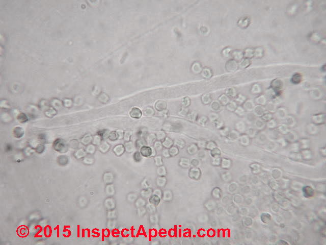
Aureobasidium pullulans fungal spore images
We find lots of the fungus spores shown above, Aureobasidium pullulans, a black yeast fungus, growing on wet or damp wood in buildings, especially on plywood roof sheathing in poorly-vented building attics.
This yeast-like fungus is also often found on caulk or damp window frames in bathrooms. Aureobasidium may be pink or black in color. More detail is at MOLD ATLAS & PARTICLES INDEX.
And we discuss this mold further at Recognize Harmless Black Mold.
B
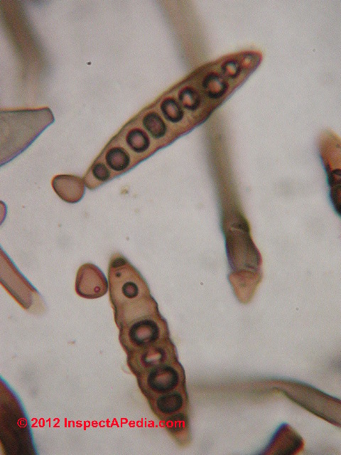
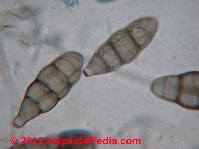
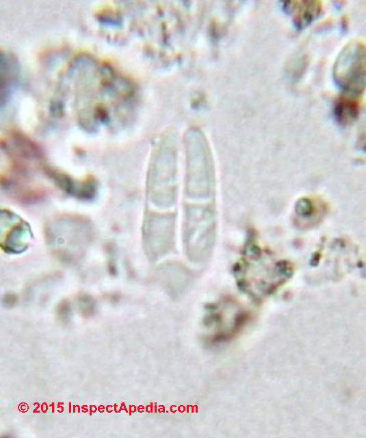
Biolaris-Drechslera sp. mold spore photographs
Lab microscopic photographs of an Bipolaris-Drechslera spores are provided below. In lab reports Bipolaris sp. and Drechslera sp. fungal spores are often grouped together as a class because of physical similarity.
C
Ceratocystis/Ophistoma - the Ophistomoid Cosmetic Black Sapstain or Bluestain Molds on Lumber

Above we show a sketch of the perithecium, ascospores, and conidia of Endoconodiphora coerulescens from the July-August 1953 issue of Mycologia Vol XLV No. 4.
Laboratory microscopic photographs of Ceratocystis/Ophistoma type mold are a bit tricky in surface samples such as collected from moldy lumber, because usually this mold is dry, often encysted, and because it is not likely to be growing on an indoor surface, the sample may lack clear identifying particles or structures.
Our photograph just above shows a fungus found under a basement stairwell that we classified as Ceratocystis/Ophistoma, and in this photograph you can see an enlarged closeup of mold fragments from that sample.
Watch out: Because this dark colored fungal growth appears "black" on wood surfaces, scaring some folks into unnecessary and costly "toxic black mold remediation" projects, we discuss the cosmetic Ceratocystis/Ophistoma mold group in detail at Recognize Harmless Black Mold.
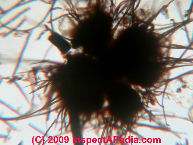
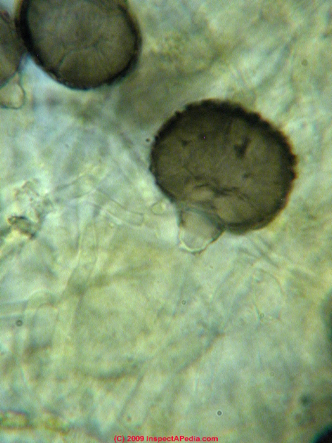
Chaetomium sp. mold spore photographs
Lab microscopic photographs of an Chaetomium spores: Chaetomium sp. (C. globosum, C. aureum, and others) are very common indoor molds found especially where drywall or other paper covered products have been wet.
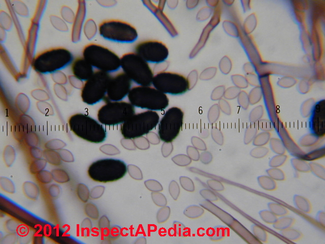
Chaetomium sp. (above) is an Ascomycete and is ubiquitous in water damaged buildings, especially on drywall paper. We find Chaetomium fungal growth often co-existing with Stachybotrys sp./ S. chartarum (photo below right - the S. chartarum are the ovate black spores)
Where Chaetomium mold growth has been found indoors in spore clusters like this it is probably appropriate to investigate the building leak history and to remain alert for the presence of other indoor mold reservoirs.
Watch out: What Chaetomium fungal spores look like in the microscope depends a lot on how they are prepared (what mountant chemicals) and the extent of spore hydration.
So Chaetomium that is not well hydrated remains "folded" to produce a center furrow that can cause it to be mistaken for Arthrinium sp. (a mistake we see in Grant Smith's execllent book of mold photos) and other molds.
Our Chaetomiium sp. photo above illustrates both hydrated and under-hydrated spores. Below we see a close-up of a few Chaetomium spores at 1200x via our Polam microscope.
Below: Chaetomium globosum under the microscope.
Warning: when dessicated these spores can fold inwards to produce a false scar.
Cladosporium sp. mold spore photographs
Microscope photographs of Cladosporium sp.: Cladosporium sp. are the most common mold spore found in outdoor air in many areas, so common in fact that Cladosporium is called "the king of molds".
The photograph of Cladosporium sp. spores in a cluster (above ) shows these spores with their proper coloration.
The Cladosporium sp. photograph at above right demonstrates the confusion in spore coloration and thus in spore identification that can occur when a novice microscopist relies too heavily on fuchsin stains for spore detection. However both photographs show the characteristic dark scars at the attachment point for these mold spores.
Our photograph just abovce shows the dominant spherical spores produced by Cladosporium sphaerospermum - another common indoor and attic/roof-sheathing mold.
Curvularia sp. mold spore photographs

Curvularia mold spores above may not be looking their best in this field photo but this is what you're likely to see at the microscope.
The Curvularia sp. belowt was in better condition, showing its attachment scar as well.
D
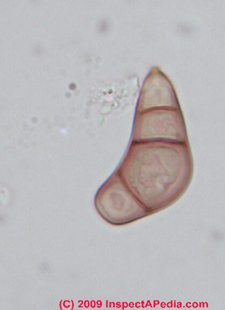
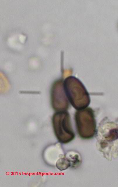
Drechslera sp. mold spore photographs
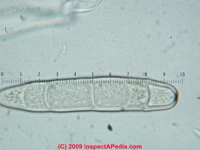
Microscope photographs of Drechslera sp. fungal spores - under the microscope Drechslera and Bipolaris mold spores are both large, segmented spores such as the member shown here, and may require additional careful examination to differentiate the two.
E
Epicoccum sp. mold spore photographs
Microscope photographs of Epicoccum sp. (E. niger)fungal spores under the microscope are large, segmented spores such as the member shown here.
F
Fuglio septica mold & mold spore spore photographs - "Dog vomit" mold.
Fuglio septica is shown under the stereoscopic microscope (less than 100x) at above left, while Fuglio septica mold spores are shown at above right.
This fungus is affectionately called "dog vomit mold" by some field investigators as when found growing outdoors on bark chips that's about what it looks like. We do not normally find this mold growing indoors.
G
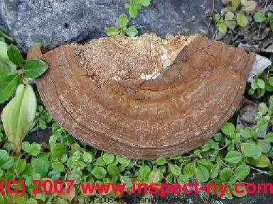
Ganoderma sp. mold spore photographs

Microscope photographs of Ganoderma basidiospores such as G. applanatum & G. tsuge dominated the air at a recent fungal spore study workshop sponsored by the University of Montreal. Here is where they were coming from.
Gonadobotryum mold spore microphotographs
Here are microscopoic photographs of Gonadobotryum sp. mold collected during a building investigation. We often find Gonadobotryum sp. mold growth present as a parasite, growing on top of other fungi, especially in the Ceratocystis/Ophistoma group on framing lumber.
The presence of Gonadobotryum sp. mold on framing lumber is probably not a health concern; it has not been reported as producing mycotoxins but we would not rule out its possible role as an allergen.
HIJKL
M
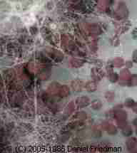
Meruliporia Incrassata - "Poria" "House Eating Fungus" Mold Microphotographs
Here are microscopoic photographs of Meruliporia incrassata mold spores collected in a building where extensive structural rot was found and "yellow mold" was visible on some of the rotting lumber.
We discuss this fungus in more detail at Meruliporia Mold Photographs.
N
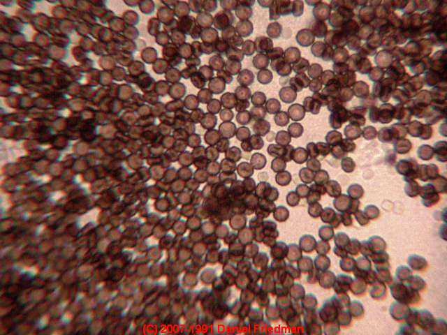
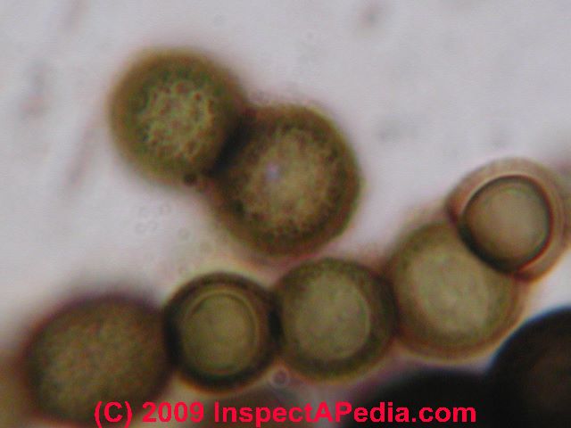
Nigrospora sp. mold spore photographs
Nigrospora sp. mold spores are often round, smooth, and black under the microscope.
It is useful to check out black round "spores" under the microscope using top lighting in order to distinguish them from paint droplets where paint has been sprayed in the building. If the round spherical objects are all smooth but their size varies, or if toplighting shows that the "spores" are white or some other color, you're probably looking at spray paint droplets, not Nigrospora sp. mold spores.
O
Oidium (Mildew) sp. mold spore photographs
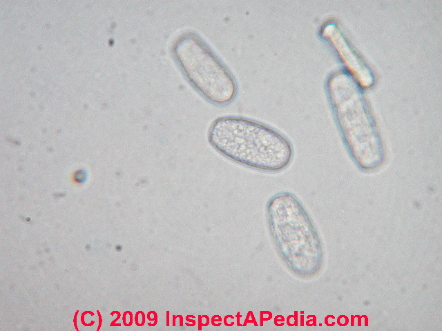
Oidium sp. is one of the most common appearances of the sub-group of molds in the mildew family.
This mold is often found in outdoor air. We do not normally find mildew growing on any indoor surface in buildings because the mildews are obligate parasites - growing only on living plants.
These spores are easy to identify by their color (none or hyaline), and their shape as well as their cellular inclusions or surface decorations visible in any sharply focused microscope at 400x or higher.
P
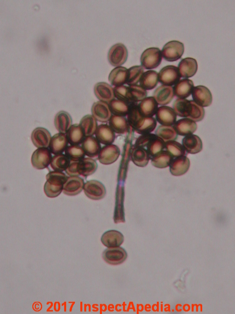
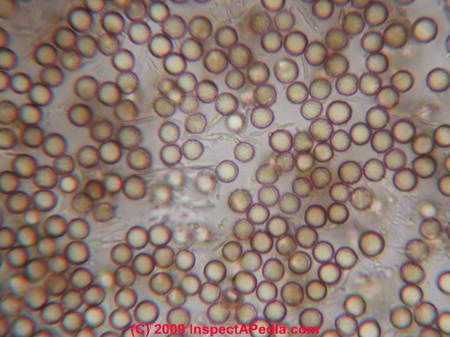
Penicillium sp. mold spore photographs
Penicillium sp. mold spores are very easy to identify when their spore producing conidiophores are collected in a surface sample (photograph at left).
But individual Penicillium sp. spores found in air or dust samples are difficult to distinguish (visually) from many Aspergillus spores as well as some other genera/species including some mold spores from a very different group, the Basidiomycetes.
A lab report of the presence of Amerospores (a generic name for unidentified small round colorless un featured spores) should not be assumed to have detected Penicillium sp.
Periconia sp. mold spore photographs
Periconia sp. mold spores are common at low levels in both indoor and outdoor air and dust samples.
Phoma sp. mold spore photographs
Phoma sp. mold spores are sometimes found indoors on building surfaces where leaks and rot damage are present.
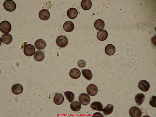
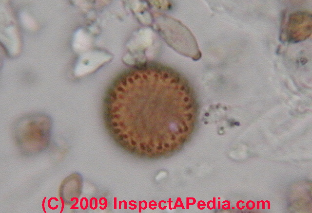
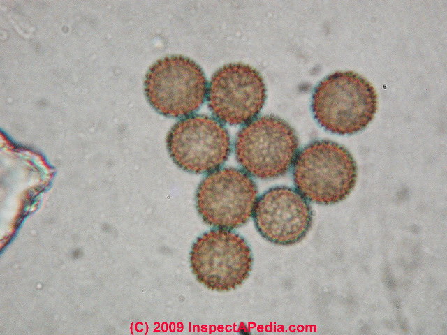
Puffball mold spore photographs
Puffball mold spores (Basidiomycetes) are common in outdoor air samples in some seasons.
The spores are easily distinguished by their little hyphal stem attachments making them look a lot like tiny balloons.
If you ever stomped on those brown dried fungal clumps (when you were a kid), sending clouds of brown dust into the air, these photos show what you were sending aloft.
Native americans used puffball spores for medicinal purposes as well, possibly as a clotting agent on wounds.
Q
...
R
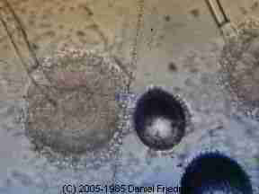
Rhizopus fungal spore & spore structure photographs - Rhizopus oryzae sporangium

At lower-left in the Rhizopus photograph above is (apparently) a dust mite fecal pellet comprised of fungal spores - Ed.
S
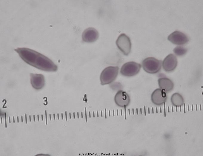
Smut spore photographs
Smut spores common in outdoor air samples, would be unusual indoors.
Sporomiella fungal spores (Ascomycetes)
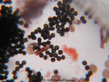
Stachybotrys sp. / Stachybotrys chartarum black mold spore photographs
Stachybotrys sp. "black mold" spore photographs under the microscope as well as on building surfaces are provided here.
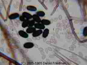
Stachybotrys chartarum mold contamination on a wall, [image]
Stachybotrys mold structure, [image]
Stachybotrys spores (with basidiospores (?), [image]
Stachybotrys mold in culture [image] has been identified (and then later questioned) as a cause of and pulmonary hemorrhage and hemosiderosis in infants. (http://gcrc.meds.cwru.edu/stachy/default.htm) While this mold has received recent media attention, many molds occur naturally outdoors and indoors.
")
Some other more common mold spores such as Penicillium and Aspergillus (see above) may cause illness or may be associated or aggravate with some types of asthma.
Stachybotrys mold, in another form, MEMNONIELLA ECHINATA [image]we've found to be particularly reactive even in small quantities. When found on building surfaces it should be removed.
Stemonitis mold & mold spore photographs

Stemonitis mold & photographs of stemonitis mold spores common growing indoors on wet oriented strand board.
T
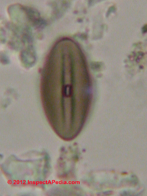
Torula sp. mold spore micro-photographs
Torula sp. fungal spores are shown in this lab photograph taken through the microscope, probably Torula herbarum.
We often encounter this mold on wet moldy or rotted plywood subfloors in buildings.
U
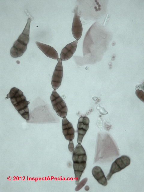
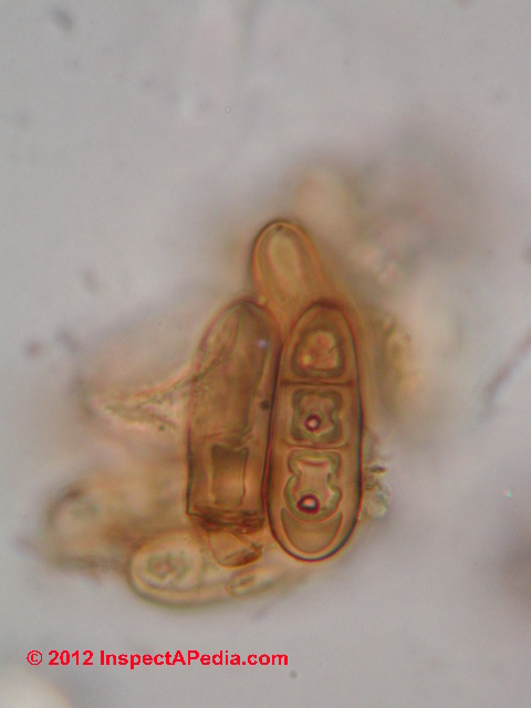
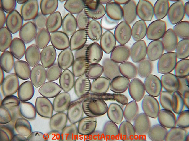
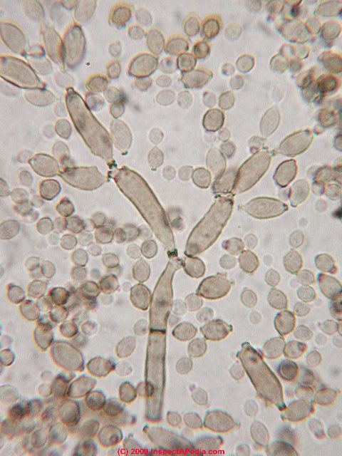
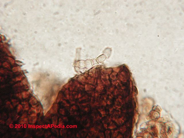
Ulocladium mold spore micro-photographs
Ulocladium sp. is often confused with similar looking versions of Stemphylium sp. and with some species of Alternaria sp. particularly as immature Alternaria spores can look like the simpler ovate cross-septated Ulocladium chartarum.
And worse there are species of Ulocladium (U. alternariae - cf Ellis) that look like (and are even named after Altenaria sp.).
The differentiation between species of Alternaria and Ulocladium is not difficult once you've been instructed by a mycologist. The "tail" you see on the Alternaria-like mold spores still attached to hyphae (photo below right) comprises the attachment point for the spore to its hyphae.
The "tail" on an Alternaria spore is at the opposite end of the mold from its attachment.
That is, an Alternaria spore is attached to its hyphae at its larger "head" end, not by its tail.
Ulocladium chartarum (below-left). Ulocladium sp. (below right).
Below are microphotographs of U. chartarum grown in culture by the author [DF].
We have almost 900,000 photomicrographs of mold spores and pollen grains - more are coming to this website. If you need to see a specific image, contact us. Content Use / Reproduction Policy - it's OK to reproduce web articles in non-commercial-use printed form only, do not make electronic copies.
MAKING ELECTRONIC COPIES OF our WEBSITE CONTENTS (such as copying our pages, content from our pages, or our graphics to another website or into email) IS EXPRESSLY PROHIBITED - this website is © protected material, all rights reserved.
V
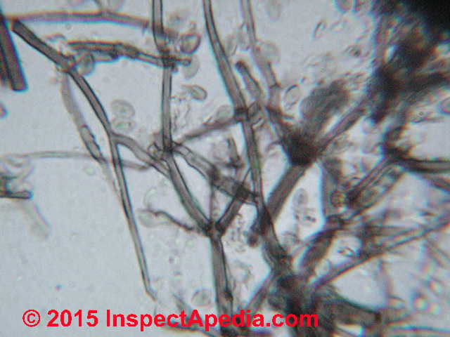
Verticillium sp. or Verticillium-like mold spore hyphae and spore photographs
W
Wallemia serbi or Wallemia-like fungal spore photographs
XY
Z
Zygophiala jamaicensis -like fungal spore photographs
...
Reader Comments, Questions & Answers About The Article Above
Below you will find questions and answers previously posted on this page at its page bottom reader comment box.
Reader Q&A - also see RECOMMENDED ARTICLES & FAQs
Question:
May I know if I can spot on cloth and wonder if that is cause by mold, what would be the test charge with microscopy identification in your side? - Ann 5/11/11
Reply:
Ann,
If you are discussing mold on clothing or a cloth curtain, in my opinion it would not be economically justified to test it unless that test were necessary as part of a wider environmental investigation. The cost of testing a tape or vacuum sample of surface mold is typically $50. - which is more than the cost of having the item professionally cleaned or in some cases just replaced.
Question: can you see mold spores under a regular light microscope? What about cellular structures & nucleii?
if you were looking under a regular light microscope at fungus, would you see spores and would you see anything inside cell such as nucleus or would you need a more powerful microscope? - Alan 5/21/2012
Reply:
Alan
Take a look at the mold spore photographs in the article above. The highest magnification we can use under most light microscopy is around 1200x, enough to see small spores down at the 1 micron level. In some cases of larger spores or more likely larger pollen grains, one can sometimes make out internal parts and components of an individual pollen grain or mold spore, but not the nucleus of an individual cell.
Question:
Our basement flooded during hurricane Irene in 2011. When we finally got power, we used a heavy industrial vac to remove the water from the basement. We moved everything into a dryer corner of the basement, and ran dehumidifiers. The entire basement seemed to dry within a matter of 2 days.
My dilemma, I never checked the boxes, cedar chest, suitcase and smaller plastic containers because we were so busy salvaging other items that were easier to move and salvage. It is 1 year and 2 months later, and we now have a generator.
In preparation for hurricane Sandy, I wanted to be prepared by cleaning and organizing the basement. While moving containers and boxes, I opened my cardboard boxes, cedar chest, suitcase and containers to see what I could keep and throw away. I was DEVASTATED!!!! There was water in the bottom of each of the containers, with some strange appearance of yellowish, greenish, and some black mold on papers and dishes.
and especially apparent on all of my shoes, scarves, mittens, boots, board games, and clothes. I barely opened the remaining containers. Instead, I slid each box & containers into large industrial size black garbage bags, and took them outside. I now have the antique cedar chest to contend to, but am not sure if I should attempt to salvage anything. In fact, I think much of the stuff in there are sweaters, blankets, dishes, and some other household items.
Is it worth opening my cedar chest to examine the items? Or would I be jeopardizing my health? I have been using rubber gloves, and masks, but am still afraid of exposure, and I am not ready to die just yet! What are the risks, and what type of solution can I use to clean (if salvageable) the cedar chest, and other personal belongings such as shoes, boots, scarves, sweaters, and gloves? Your response is much appreciated! Thank you! - MoMo 10/31/2012
Reader further comment:\
I took pictures of the things that were precious to me; gave thanks and praise to God for the blessings that I had and still have, and then threw everything out!
Reply:
MoMO
I apologize for the delay in response - as you might imagine, following Hurricane Sandy in 2012, we've been swamped with queries including some from folks who are homeless or ill. You were asking about salvaging materials that were flooded by Hurricane Irene more than a year ago, back in 2011.
At MOLD ACTION GUIDE - WHAT TO DO ABOUT MOLD we provide procedures for cleaning up a mold problem, and
at EMERGENCY RESPONSE, IAQ, GAS, MOLD as well as
at DISASTERS: BUILDING INSPECTION & REPAIR we describe procedures for handling building flooding following a hurricane, storm surge, sewer backup or similar problem.
It appears to me that the remediation you performed in your basement after Hurricane Irene in 2011 may have been seriously inadequate, in particular, if you simply vaccumed up water and ran dehumidifiers without removing wet drywall, insulation, or soaked materials and stored items. Only by removing wet moldy debris can we move towards a successful cleanup;
I agree with your step of bagging and moving wet or moldy items out of the building for inspection, salvage, or disposal beginning outdoors.
Generally, after having been soaked and perhaps moldy since 2011, the sorts of items that can be easily salvaged are hard-surfaced materials such as metal, ceramics, some plastics.
What are beyond easy salvage are papers, leather clothing, shoes, etc. - those are usually beyond economical repair or cleaning. Some fabrics & other clothing, if valuable enough (the item is worth more than the cost to clean it) , can be salvaged by laundering and/or drycleaning, though if the materials have been soaked and moldy for more than a year, it may be difficult to completely remove odors without expert (and more costly) treatment.
At BOOK / DOCUMENT MOLD & WATER DAMAGE RESTORATION you can read about how we deal with books or papers that people want to save after flooding.
Basically, if you have moldy papers that you need to save but cannot afford to clean, try drying them in sunlight (some folks try a microwave located outdoors), followed by gentle wiping and then store these modly items in a dry air-tight plastic box. The items will not have been cleaned and should not be handled without appropriate precautions, but they can at least be retained.
As for jeapordizing your health - if you are not someone at extra risk (asthmatic, immune compromised, elderly, etc) and if you wear appropriate protective gear you can open those items stored out of the home for inspection and sorting into trash and salvage piles.
Thank you for the question.
...
Continue reading at MOLD in the PETRI DISH, PHOTOS or select a topic from the closely-related articles below, or see the complete ARTICLE INDEX.
Or see these
Directories of atlases or indices of building mold
- BLACK MOLD, HARMLESS - Photos of of often recognizable, usually harmless or cosmetic black mold on wood
- MOLD ATLAS & PARTICLES INDEX, Pathogens, Allergens and Other Indoor Particles - actual and possible medical health effects of Mold (separate online document)
- MOLD FREQUENCY in BUILDINGS - Table of what mold genera/species are frequently found in indoor mold tests.
- MOLD GROWTH ON SURFACES, PHOTOS - Photos of what mold looks like in buildings on more than 100 surfaces, organized by mold growth on various kinds of building surfaces and contents or items found in buildings. Mold in situ.
- MOLD GROWTH on SURFACES, TABLE OF - Summary table of what mold genera/species are frequently found on various building surfaces and materials
- MOLD BY MICROSCOPE - Mold under the microscope - photo identification of the most common indoor molds found in buildings
- MOLD in the PETRI DISH, PHOTOS - what mold looks like on culture plates or in culture-type mold test kits
- MOLD RELATED ILLNESS SYMPTOMS - Mold Related Illness: Index of Symptoms. Readers should not rely on this document for medical diagnosis and instead should consult with their physician or with a specialist such as a medical toxicologist
Articles on What Mold Looks Like
- LAB PROCEDURES MICROSCOPE TECHNIQUES
- MOLD APPEARANCE - WHAT MOLD LOOKS LIKE
- BLACK MOLD IDENTIFICATION
- BLACK MOLD, HARMLESS
- BROWN MOLD PHOTOS
- COSMETIC MOLD, RECOGNIZE
- GREEN MOLD PHOTOS
- INVISIBLE MOLD
- LIGHT COLORED MOLD
- MERULIPORIA HOUSE EATING FUNGUS
- MERULIPORIA FUNGUS & TERMITE INTERACTIONS
- PORIA DAMAGE PREVENTION
- MILDEW ERRORS, IT's MOLD
- MILDEW PHOTOGRAPHS
- MOLD APPEARANCE on VARIOUS SURFACES
- MOLD ATLAS & PARTICLES INDEX
- MOLD GROWTH on SURFACES, PHOTOS
- MOLD on DIRT FLOORS
- MOLD on DIRT FAQs
- MOLD by MICROSCOPE
- MOLD in the PETRI DISH, PHOTOS
- RED MOLD PHOTOS
- YELLOW MOLD PHOTOS
- WHITE MOLD PHOTOS
- MOLD APPEARANCE - STUFF THAT IS NOT MOLD
Suggested citation for this web page
MOLD by MICROSCOPE at InspectApedia.com - online encyclopedia of building & environmental inspection, testing, diagnosis, repair, & problem prevention advice.
Or see this
INDEX to RELATED ARTICLES: ARTICLE INDEX to MOLD CONTAMINATION & REMEDIATION
Or use the SEARCH BOX found below to Ask a Question or Search InspectApedia
Ask a Question or Search InspectApedia
Try the search box just below, or if you prefer, post a question or comment in the Comments box below and we will respond promptly.
Search the InspectApedia website
Note: appearance of your Comment below may be delayed: if your comment contains an image, photograph, web link, or text that looks to the software as if it might be a web link, your posting will appear after it has been approved by a moderator. Apologies for the delay.
Only one image can be added per comment but you can post as many comments, and therefore images, as you like.
You will not receive a notification when a response to your question has been posted.
Please bookmark this page to make it easy for you to check back for our response.
Comments


IF above you see "Comment Form is loading comments..." then COMMENT BOX - countable.ca / bawkbox.com IS NOT WORKING.
In any case you are welcome to send an email directly to us at InspectApedia.com at editor@inspectApedia.com
We'll reply to you directly. Please help us help you by noting, in your email, the URL of the InspectApedia page where you wanted to comment.
Citations & References
In addition to any citations in the article above, a full list is available on request.
- ENVIRONMENTAL HEALTH & INVESTIGATION BIBLIOGRAPHY - our technical library on indoor air quality inspection, testing, laboratory procedures, forensic microscopy, etc.
- In addition to citations & references found in this article, see the research citations given at the end of the related articles found at our suggested
CONTINUE READING or RECOMMENDED ARTICLES.
- Carson, Dunlop & Associates Ltd., 120 Carlton Street Suite 407, Toronto ON M5A 4K2. Tel: (416) 964-9415 1-800-268-7070 Email: info@carsondunlop.com. Alan Carson is a past president of ASHI, the American Society of Home Inspectors.
Thanks to Alan Carson and Bob Dunlop, for permission for InspectAPedia to use text excerpts from The HOME REFERENCE BOOK - the Encyclopedia of Homes and to use illustrations from The ILLUSTRATED HOME .
Carson Dunlop Associates provides extensive home inspection education and report writing material. In gratitude we provide links to tsome Carson Dunlop Associates products and services.
 | HOME | ABOUT | ASK a QUESTION | CONTACT | CONTENT USE POLICY | DESCRIPTION | POLICIES | PRIVACY | |
| © 2025 - 1985 Publisher InspectApedia.com - Daniel Friedman | |||||||||