
InspectAPedia® FREE Encyclopedia of Building & Environmental Construction, Diagnosis, Maintenance & Repair |
Question? Just ask us! InspectAPedia
|
 Asbestos under the microscope
- POST a QUESTION or COMMENT about how asbestos is identified in various materials, asbestos identification microscopy, & what asbestos looks like microscopically
Asbestos particles examined by microscope.
Photographs provided on this page illustrate what asbestos fibers or fragments may look like under the polarized light microscope.
In this article we provide photographs and descriptive text of asbestos insulation and other asbestos-containing products to permit identification of definite, probable, or possible asbestos materials in buildings.
This document assists forensic investigators, laboratories, building buyers, owners or inspectors who need to identify asbestos materials (or probable asbestos) in buildings by simple visual inspection and confirmed by asbestos test lab or forensic microscopy lab examination.
InspectAPedia tolerates no conflicts of interest. We have no relationship with advertisers, products, or services discussed at this website.
Asbestos under the microscope: Micro photographs of Asbestos
 While an expert lab test using polarized light microscopy may be needed to identify the specific type of
asbestos fiber, or to identify the presence of asbestos in air or dust samples, many asbestos containing building products
not only are obvious and easy to recognize, but since there were not other look-alike products that were not asbestos, a visual identification of this material can be virtually a certainty in many cases.
While an expert lab test using polarized light microscopy may be needed to identify the specific type of
asbestos fiber, or to identify the presence of asbestos in air or dust samples, many asbestos containing building products
not only are obvious and easy to recognize, but since there were not other look-alike products that were not asbestos, a visual identification of this material can be virtually a certainty in many cases.
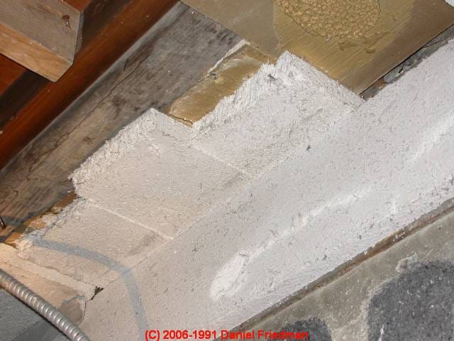
Our photo (left) illustrates pure tremolite asbestos used as fireproofing on a building ceiling in New York. Our forensic lab photographs of tremolite asbestos (below) were collected from that ceiling. More about this building can be read
at ASBESTOS FIREPROOFING SPRAY-ON COATINGS.
Also
see ASBESTOS DUCTS, HVAC a field identification guide to
visual detection of asbestos in and on heating and cooling system ducts and flue vents,
and see Micro-Photographs of DUST from the WORLD TRADE CENTER COLLAPSE following the 9/11/2001 attack. Links to U.S. government and other authoritative research and advice are included.
PLM (Polarized Light Microscopy) remains the most commonly or widely used method for identifying asbestos: with proper training, equipment, a polarized light microscope, (ideally sporting one or more central stop dispersion staining lenses) and the proper immersion oil in the right range of refractive index, this is quick, efficient, and easy.
A second, widely used means of identification of asbestos in material samples is the use of SEM (Scanning Electron Microscopy) or TEM (Transmission Electron Microscopy) approaches using a directed beam of electrons, an X-ray or EDX (energy-dispersive X-rays) beam, and proper computer software and hardware in support. This approach has the benefit of easier identification of different types of asbestos, though Crone also showed that asbestos types could also be identified by his methodology.
REMs are of course a higher resolution technology than a PLM that is generally limited to particles down to about 1u at 1200x or smaller particles at 1920x magnification (using our POLAM Russian-built PLM).
TEM has still higher resolution. However in our OPINION as well as that of experts, PLM is completely adequate for identifying asbestos in bulk, air, or dust samples.
 ...
... 
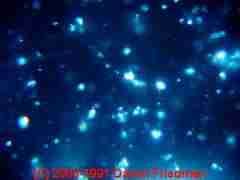
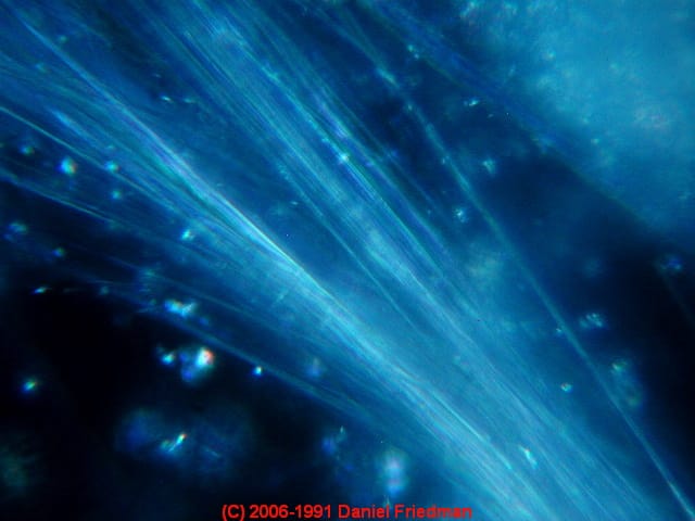
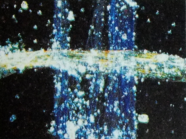
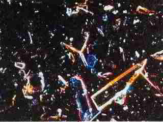
Above are two photographs showing what a sample of asbestos ceiling fireproofing (tremolite asbestos) looks like in our lab microscope using polarized light microscopy (PLM).
Notice that in the first photo you see long very thin multi-fibrous filaments - asbestiform tremolite.
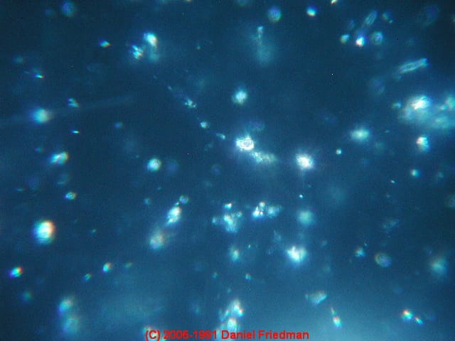
Each filament is less than one micron in diameter. In the second photograph you'll observe non-fibrous granular particles, many less than one micron in diameter as well - non-asbestiform tremolite. [Crone]
This asbestos sample was collected from slabs of nearly pure tremolite asbestos which was
used
as FIREPROOFING ASBESTOS SPRAY-ON in a commercial building.
Crone illustrated that tremolite asbestos (as well as some other forms of asbestos) occur in both fibrous and non-fibrous form.
Comparing the photo at below left (tremolite in fibrous form) by Crone to ours (above right) that shows fewer small non-fibrous particles, but a clear bundle of ultra-fine sub-micron (in width) fibers.
 ...
... 
Asbestos in Boiler & Pipe Lagging Insulation used on a Wall
Reader Question: Can You Identify This Dense Spongy Heavy Insulation Chrysotile Asbestos
Thank you for the follow-up to my post on the website regarding the strange insulation uncovered at my 1863 house in southeastern Indiana. You may recall that the material is dense, spongy, and very heavy.

I sent a sample for asbestos testing which came back positive for 10% Chrysotile asbestos. You mentioned in your reply that you would like to examine it in your lab. I retained a sample, double bagged and ready to go. I’d be grateful and very interested to know the results.
I am also interested to know the makeup of the material. We already know that it contains asbestos but it will be very interesting to know what was used at the time.
(I can also include a piece of the paper backing for your examination.) Please advise. - Anonymous by private email 2022/11/08

P.S. do you want a small piece of the backing paper as well? Perhaps it will help in identifying the product manufacturer
Moderator Reply: Chrysotile asbestos appears mixed with plaster or gypsum as was often used to form a paste or coating on boilers and pipe elbows, here plastered on a wall.
That sure looks as if someone used an asbestos and plaster mix that was applied to wood lath as part of a building wall, perhaps as an effort at fire resistance.
In your first photo, if we click to enlarge and zoom on the image we can see nail holes and a few nails that appear to show where wood lath was installed.
It would be helpful to know the exact location and context of the asbestos insulation in your photos.
I would not attempt to duplicate a certified asbestos test lab's work but I am interested in characterizing the material under the microscope and transmitted into polarized light and I'll be glad to share those results.
The best way is to mail a small sample, I don't need nor want more than a square inch, enclose it in a freezer type Ziploc bag and put that inside of another bag and then mail that to me in a suitable envelope as follows ... [details omitted here - Ed.]
A photo of the backer or surrounding paper or packaging, in particular if you come across any stampings, letters, logos, etc. is particularly helpful.
Watch out: note to readers: don't send a large volume of unknown material like this to your test lab. Large volumes are more difficult to handle and may be unsafe. Some forensic microscopy labs will simply throw away such samples or may charge extra for handling them.
A single cubic inch of representative material is plenty.
Asbestos Insulation Under the Microscope
I'm just back in the lab today and have looked at the (way too much) sample material you sent.
 ...
... 
I examined a small sample of the material both in the stereo microscope and in the high powered forensic microscope, using several preparing chemicals, and at magnifications up to 1200x with transmitted, reflected, and polarized light.
If you can send along some sharp photos of the paper backer you mentioned, particularly to include any text or logos, that would be valuable.

Above: The insulating material under the stereo microscope looks as if it includes a combination of fibers and filler, dominated by the filler material. Here we are using top lighting and you can see the characteristic white color of the material.
Below: Under the Polam forensic microscope: the asbestos lagging insulation under the microscope at low power, using transmitted light. Notice that we have a synthetic fiber and another cellulosic fiber at the right side of the material, useful for scale.

When I used a non-acidic slide prep chemical (triacetin) the powder formed a mush that was diagnostic but not so nice under the microscope.
When I used an acidic slide preparation chemical (Lactic acid) the material foamed and much of it dissolved: something I've observed many times with plaster dust and drywall dust.
What was left included, however, both fibers and very small granular particles that under polarized light look to be forms of asbestos, perhaps more than one form. Here we see what looks like a fabric fiber that gives great scale for those few long asbestos fibers and the smaller fragments. That image is just below.

Above, still with transmitted light, we see a combination of filler and some very thin fibers - at about 100x using the Polam microscope.
Now in the two photos below we compare a view of the same sample in transmitted light and polarized light at higher magnification.
 ...
... 
Below: next we see a magnified view of one of those very thin fibers, now at 1200x using polarized light. The result is characteristic of asbestos fibers, made up of multiple strands each down under 1u in thickness.

Below: This photo provides a combination of asbestos fibers and granular particles that may form asbestos "shorts" or filler.
Most of the gypsum-like filler has been dissolved by use of an acidic mountant.

Now as a possible source of that plaster-asbestos material, can you tell me (and show me photos of) a steam heating system in that home?
My opinion is that the "insulation" that you sent is a mix of a lot of asbestos, including asbestos fibers and asbestos "shorts" that show up as powder, mixed with what is most likely gypsum or plaster base.
This could well be from someone's supply of asbestos lagging that was used to form a paste that was in turn used to insulate heating boilers as well as pipe elbows. The sample also contained a few extraneous fibers, perhaps synthetic fabric, such as the red fiber shown in our earlier low magnification photo.
I'd also like to see a copy of the asbestos test lab report that you received showing 10% chrysotile asbestos. That's reasonable but I think there may be more asbestos in other forms that was harder to identify, depending on the lab methodology.
Asbestos Identification Resources
 ...
... 
- EPA Guidance for Controlling Asbestos-Containing Materials in buildings, NIAST, National Institute on Abatement Sciences & Technology, [republishing EPA public documents] 1985 ed., Exposure Evaluation Division, Office of Toxic Substances, Office of Pesticides and Toxic Substances, U.S. Environmental Protection Agency, Washington,D.C. 20460
- ASBESTOS IN YOUR HOME U.S. EPA, Exposure Evaluation Division, Office of Toxic Substances, Office of Pesticides and Toxic Substances, U.S. Environmental Protection Agency, Washington,D.C. 20460
- Asbestos products and their history and use in various building materials such as asphalt and vinyl flooring includes discussion which draws on ASBESTOS, ITS INDUSTRIAL APPLICATIONS, ROSATO 1959, D.V. Rosato, engineering consultant, Newton, MA, Reinhold Publishing, 1959 Library of Congress Catalog Card No.: 59-12535 (out of print, text and images available at InspectAPedia.com).
- De Stefano, L., and F. De Luca. "SEM Quantitative Determination of Asbestos in Bulk Materials." MICROSCOPY AND ANALYSIS (2002): 13-16.
- McCrone, Walter C., Asbestos Identification, [book for sale at Amazon] McCrone Research Institute, Chicago, IL.1987 ISBN 0-904962-11-3. Dr. McCrone literally "wrote the book" on asbestos identification procedures which formed the basis for current work by asbestos identification laboratories.
- McCrone, Walter C., Lucy B. McCrone, John Gustav Delly, "Polarized Light Microscopy", McCrone Research Institute, Chicago (2003 Fourteenth printing) , original copy 1984, ISBN 0-250-40262-9.
Preface Excerpt:
This volume is in reality a recent edition of a course manual first prepared by Walter C. McCrone and Marven Salzenstein in 1959. [The first] hard-cover edition was printed in 1977.
Disclosure: InspectApedia's editor/author took this course as a student at McResearch Institute in 2003.- Ed. - Stanton, .F., et al., National Bureau of Standards Special Publication 506: 143-151
- Pott, F., Staub-Reinhalf Luft 38, 486-490 (1978) cited by McCrone
Reader Comments, Questions & Answers About The Article Above
Below you will find questions and answers previously posted on this page at its page bottom reader comment box.
Reader Q&A - also see RECOMMENDED ARTICLES & FAQs
Question: asbestos fibers in the nose?
(Nov 2, 2014) Doreen said:
If asbestos fibers get trapped in nose and sinus that are open and/or infected,can serious harm occur? If so, in what way? I believe I may be being exposed and am suffering some severe effects.
Reply:
Doreen:
Large fibers of asbestos, cloth, lint, etc. that are "trapped' in the nose are normally secreted, e.g. when you blow your nose or clean it.
Large fibers of any material that are breathed into the lungs are typically exhaled or coughed out or lodged high in the lung.
Small particles of any material, including asbestos fibers, are more dangerous as they are easily breathed deeply into the lungs and are not so easily expelled. Worse, smokers have an 80 times greater risk of lung cancer related to radon exposure and I suspect similar hazards apply to inhalation of other small particles by smokers, including asbestos or silica.
Doreen you should take your concerns to your doctor.
...
Continue reading at ASBESTOS TESTING LAB LIST or select a topic from the closely-related articles below, or see the complete ARTICLE INDEX.
Or see these
Recommended Articles
- ASBESTOS in TALCUM POWDER
- FIBER & HAIR IDENTIFICATION
- FORENSIC INVESTIGATION of BUILDINGS
- FORENSIC & IAQ LAB MICROSCOPE TECHNIQUES
Suggested citation for this web page
ASBESTOS UNDER the MICROSCOPE at InspectApedia.com - online encyclopedia of building & environmental inspection, testing, diagnosis, repair, & problem prevention advice.
Or see this
INDEX to RELATED ARTICLES: ARTICLE INDEX to ASBESTOS HAZARDS
Or use the SEARCH BOX found below to Ask a Question or Search InspectApedia
Or See
INDEX to RELATED ARTICLES: ARTICLE INDEX to BUILDING FORENSICS
Or use the SEARCH BOX found below to Ask a Question or Search InspectApedia
Ask a Question or Search InspectApedia
Try the search box just below, or if you prefer, post a question or comment in the Comments box below and we will respond promptly.
Search the InspectApedia website
Note: appearance of your Comment below may be delayed: if your comment contains an image, photograph, web link, or text that looks to the software as if it might be a web link, your posting will appear after it has been approved by a moderator. Apologies for the delay.
Only one image can be added per comment but you can post as many comments, and therefore images, as you like.
You will not receive a notification when a response to your question has been posted.
Please bookmark this page to make it easy for you to check back for our response.
Our Comment Box is provided by Countable Web Productions countable.ca
Citations & References
In addition to any citations in the article above, a full list is available on request.
- 3/07: thanks to Gary Randolph, Ounce of Prevention Home Inspection, LLC Buffalo, NY, for attentive reading and editing suggestions. Mr. Randolph can be reached in Buffalo, NY, at (716) 636-3865 or email: gary@ouncehome.com
- 06/07: thanks for photographs of transite asbestos heating ducts, courtesy of Thomas Hauswirth, Managing Member of Beacon Fine Home Inspections, LLC and (in 2007) Vice President, Connecticut Association of Home Inspectors Ph. 860-526-3355 Fax 860-526-2942 beaconinspections@sbcglobal.net
- June 1997 - Window Putty - OSHA case cites contractor for asbestos exposure during removal of window putty http://www.osha.gov/pls/oshaweb/owadisp.show_document?p_table=NEWS_RELEASES&p_id=1091
- "Handling Asbestos-Containing roofing material - an update", Carl Good, NRCA Associate Executive Director, Professional Roofing, February 1992, p. 38-43
- In addition to citations & references found in this article, see the research citations given at the end of the related articles found at our suggested
CONTINUE READING or RECOMMENDED ARTICLES.
- Carson, Dunlop & Associates Ltd., 120 Carlton Street Suite 407, Toronto ON M5A 4K2. Tel: (416) 964-9415 1-800-268-7070 Email: info@carsondunlop.com. Alan Carson is a past president of ASHI, the American Society of Home Inspectors.
Thanks to Alan Carson and Bob Dunlop, for permission for InspectAPedia to use text excerpts from The HOME REFERENCE BOOK - the Encyclopedia of Homes and to use illustrations from The ILLUSTRATED HOME .
Carson Dunlop Associates provides extensive home inspection education and report writing material. In gratitude we provide links to tsome Carson Dunlop Associates products and services.
 | HOME | ABOUT | ASK a QUESTION | CONTACT | CONTENT USE POLICY | DESCRIPTION | POLICIES | PRIVACY | |
| © 2024 - 1985 Publisher InspectApedia.com - Daniel Friedman | |||||||||